I now routinely use ‘dimple slides’ – glass microscope slides with a well in them. A glass cover-slip can be put over the drop of moss squeeze water. The moss squeeze sample can then be observed under a light microscope without crushing the organisms that are within it (Figure 1. A). If you don’t have dimple slides here are some alternative suggestions.
If you place a couple of drops of moss squeeze water onto a flat glass microscope slide, it forms a neat dome due to the cohesive behaviour of water molecules. Inside that dome will be organisms and debris that you have taken from the moss sample. If you were to place this under a light microscope, you may see some of the contents, but focusing is difficult and there are issues with light reflecting from the dome surface. In addition, you could contaminate the objectives if you were to touch them on the water drop. It is better to look at a flattened drop, but without squashing the organisms within it.
One way to do this is to invert the slide, so the water droplet hangs on the underside of the slide (Figure 1. B). The sample is observed through the glass slide, through the flat side of the droplet. This removes distortions and reflections and protects the objectives. The disadvantage is that the drop doesn’t always behave, it takes a bit of practice. Also if above the microscope or a while, the water can evaporate quite quickly.
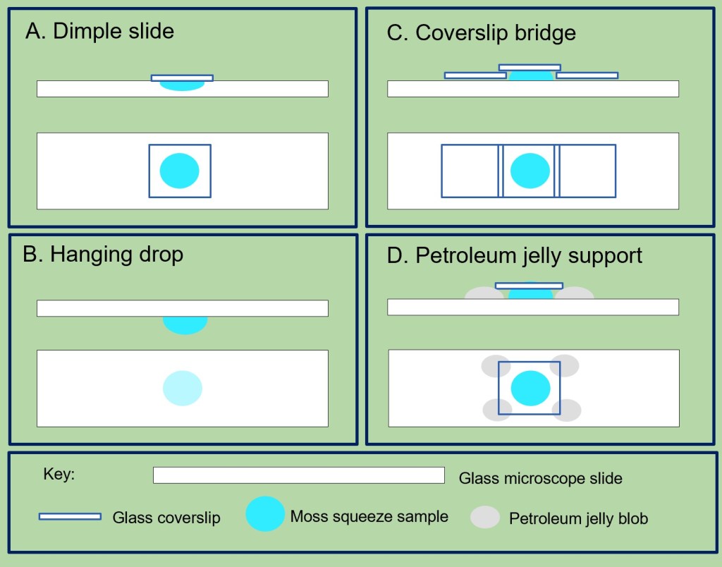
If you have glass cover-slips available, but without a dimple slide, you can try the final two methods. One method is making ‘cover-slip bridge’ (Figure 1. C). Place a couple of drops of the moss squeeze sample into the centre of a flat glass microscope slide. Place one cover-slip on each side of the drop, then place a final cover-slip over the drop so it rests on the two cover-slips, like a bridge. This will flatten the droplet without harming the organisms. If cover-slips are in short supply, using a dab of petroleum jelly on each corner (Figure 1. D), holds the slip above the droplet in a similar way to the bridge technique. Care has to be taken not to get the jelly on the microscope objectives.
What methods do you use?
Suppliers of the ‘dimple’ slides, also known as ‘cavity’ or ‘concave’ microscope slides include: Amazon, Philip Harris, and other science equipment suppliers. Depending on quality and quantity bought, they around 25p – £1.50 each. They can be reused.
